BIOcean5D is dedicated to exploring marine life and understanding how it changes across space, time and human impact. Valuable insights can be gained by studying how the morphology of microorganisms varies across different environments and conditions. To support this effort, and help fill the marine biodiversity knowledge gap, fluorescence microscopy images have been generated of hundreds of plankton samples collected during the TREC – Tara Europa expedition.
Tina Wiegand, Senior Light Microscopy Specialist at EMBL, explains: “These subcellular images allow us to visualise the state of microorganisms and quantify structural parameters such as size and volume. They also help identify organisms with high photosynthetic pigment content, such as chlorophyll, and reveal potential symbiotic relationships.”
A Unique Expedition
TREC – Tara Europa was a unique and unprecedented scientific expedition that collected samples along the entire European coastline to explore the biodiversity of coastal ecosystems. At several key locations along the expedition route – Roscoff, Tallinn, Kristineberg, Bilbao, Porto, Barcelona, Naples and Athens – parallel sampling was carried out both at sea by the Tara schooner and across the land-sea interface, including soils, sediments and shallow waters, supported by EMBL’s Advanced Mobile Laboratory (AML) on site. Tina describes these coordinated stopovers as “an extraordinary collaborative effort: scientists from different disciplines and institutions worked closely together with local researchers, and we also shared our work with the public through outreach and engagement activities.”
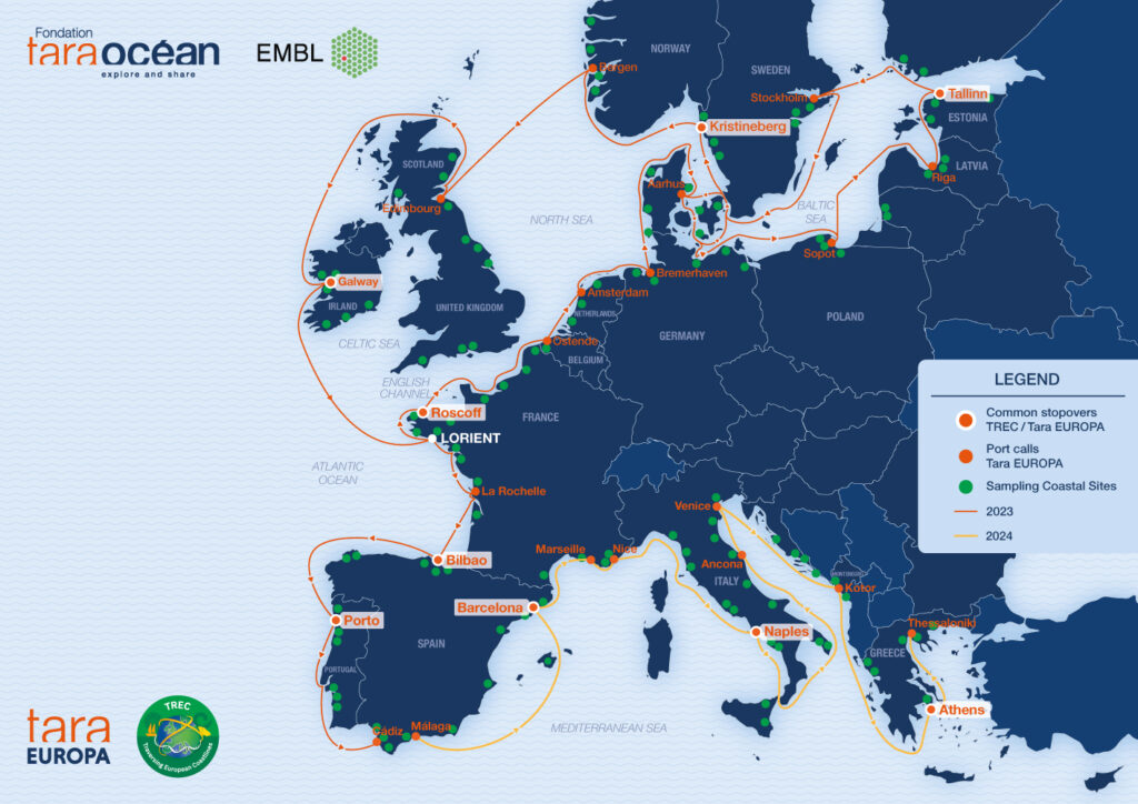 Map of the TREC – Tara Europa expedition. Credit: Tara Ocean Foundation.
Capturing Life in Real Time
From each set of plankton samples collected at these sites, a subsample was systematically analysed immediately in the AML. “The AML brings sophisticated technology directly to where scientists need it, making advanced research possible in field settings where such capabilities are not usually available,” explains Niko Leisch, Head of Mobile Laboratories Core Facility at EMBL. “Imaging live samples on site allowed us to observe cellular behaviour directly and avoid artefacts introduced by chemical fixation: morphology remains unaltered, pigments stay intact and dynamic processes can be monitored in real time,” continues Tina.
There are, however, limitations. ”Certain organisms actively move away from light or the adhesive surfaces used for imaging, making them difficult to capture. The resolution is also limited because high laser power can damage or kill the organisms.” Despite these constraints, live microscopy provided a valuable overview, including a near real-time estimation of biodiversity, abundance and health of the plankton sampled, that informed subsequent experiments and imaging strategies.
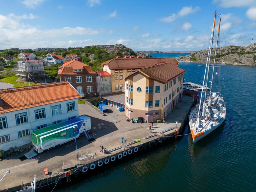 EMBL’s Advanced Mobile Laboratory located on site with the Tara schooner enabling parallel sampling in Kristineberg, Sweden in July 2023 during the TREC – Tara Europa expedition. Credit: Niko Leisch/EMBL.
Building an Open Resource
The complete microscopy dataset of more than 350 live samples – approximately 17,000 images, including over 600,000 individual objects – is now publicly available through the BioImage Archive, with segmented versions available via EcoTaxa.
“The data from days when parallel sampling was carried out is particularly valuable as the live observations can be linked to the extensive contextual and environmental data generated by scientists aboard the Tara schooner,” explains Niko. Indeed, researchers within the TREC – Tara Europa and BIOcean5D projects are already connecting microscopy data with other datasets. Tina highlights, for example, that “scientists are using the data to identify locations where specific symbiotic organisms occur, such as dinoflagellates hosting cyanobacterial endosymbionts.”
Open access also enables researchers worldwide to use the dataset for their own research questions and potentially uncover patterns, features and relationships not yet even considered. To support this, considerable work has gone into rigorously documenting methods and metadata (available via BioImage Archive) to ensure that the dataset is reusable and compatible with approaches such as artificial intelligence.
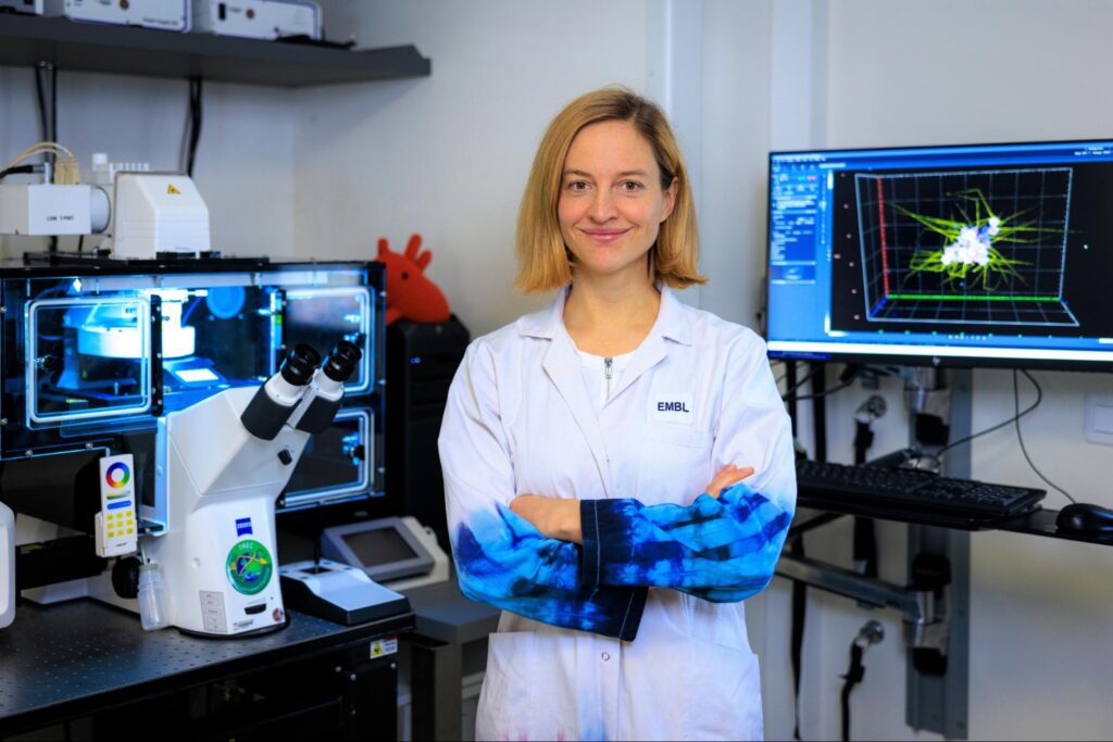 Tina Wiegand, Light Microscopy Specialist at EMBL, working inside the EMBL Advanced Mobile Laboratory to image samples collected during the TREC – Tara Europa expedition. Credit: Massimo del Prete/EMBL.
Fixed Samples for a Closer Look
Plankton samples collected during the expedition that were not imaged live were fixed on site. Following the expedition, work began to systematically stain and image these samples.
All fixed samples are first stained using a standardised protocol that enables the visualisation of DNA, lipid storage and, where present, external skeletal structures. The development of an automated fluorescence confocal microscopy workflow has significantly accelerated imaging acquisition, optimising both high-throughput and image quality. Objects of interest are first detected in overview scans, after which imaging parameters are automatically adjusted to acquire high-resolution images of selected targets. This workflow enables approximately 500 organisms to be imaged overnight with consistent image quality and an unbiased selection process.
Once completed, the microscopy dataset of fixed samples will also be made openly available. “These images provide a more comprehensive representation of the community. The use of additional fluorescent dyes, combined with higher-resolution imaging, enables the visualisation of both the cellular structure and internal organisation of these organisms,” explains Tina.
Looking Ahead
For Tina, the project has been an exceptional learning experience. “Exploring these samples has provided the opportunity to learn more about marine organisms and symbiosis, complementing my previous training in chemistry and molecular biology. I’ve also gained valuable experience in image analysis, data management and the challenges of generating and sharing such a large-scale dataset.”
The success of this work reflects the remarkable collaboration between colleagues at EMBL and across the BIOcean5D and TREC – Tara Europa projects. The resulting resource supports not only current research into marine biodiversity but — through its open availability — also creates opportunities for future discoveries and new scientific insights.